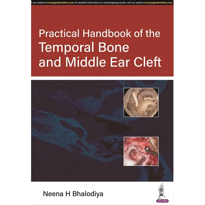
Practical Handbook of the Temporal Bone and Middle Ear Cleft
(下單前請先用官方line詢問庫存)
Quick Overview
This book consists of 6 chapters. The first 5 chapter covers the temporal bone, middle ear cavity, middle ear compartment, facial nerve, and Eustachian tube. The last chapter is exclusively on the radiology of the temporal bone. All the chapters are provided with realistic figures and CT scan pictures with fine illustrations. Flowcharts are also used where required for better understanding.
Target AudienceIt will be extremely important for budding ear, nose, and throat (ENT) surgeons and fellow ENTs.